Ophthalmoscope Optic Nerve . Scans can detect small nerve fiber layer changes of the optic nerve at the micron level. fundoscopic / ophthalmoscopic exam. click to learn how to examine cn ii (optic nerve) using techniques like visual acuity testing, color perception, assessing visual fields and accommodation! Visualization of the retina can provide lots of information about a medical diagnosis. in the procedure, one looks at structures lying in the innermost aspect of the globe, collectively known as the eyegrounds: ophthalmoscopy (also called fundoscopy or funduscopy) is an exam your doctor, optometrist, or ophthalmologist uses. the direct ophthalmoscope allows you to look into the back of the eye to look at the health of the retina, optic nerve, vasculature and vitreous humor. Retina, retinal blood vessels, optic nerve.
from depositphotos.com
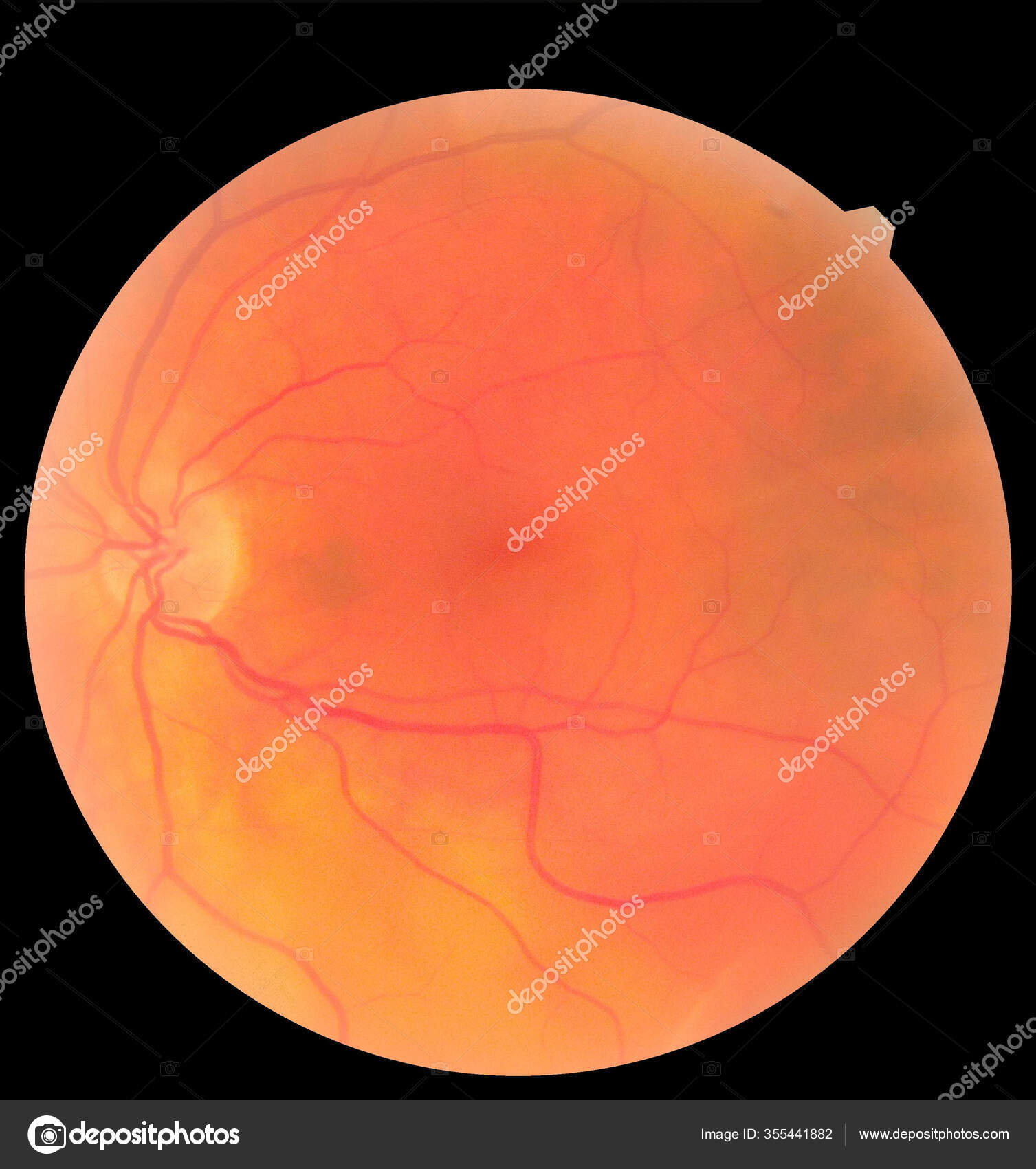
Visualization of the retina can provide lots of information about a medical diagnosis. the direct ophthalmoscope allows you to look into the back of the eye to look at the health of the retina, optic nerve, vasculature and vitreous humor. Scans can detect small nerve fiber layer changes of the optic nerve at the micron level. Retina, retinal blood vessels, optic nerve. ophthalmoscopy (also called fundoscopy or funduscopy) is an exam your doctor, optometrist, or ophthalmologist uses. in the procedure, one looks at structures lying in the innermost aspect of the globe, collectively known as the eyegrounds: fundoscopic / ophthalmoscopic exam. click to learn how to examine cn ii (optic nerve) using techniques like visual acuity testing, color perception, assessing visual fields and accommodation!
Ophthalmic image detailing the retina and optic nerve inside a healthy human eye. Health
Ophthalmoscope Optic Nerve click to learn how to examine cn ii (optic nerve) using techniques like visual acuity testing, color perception, assessing visual fields and accommodation! fundoscopic / ophthalmoscopic exam. in the procedure, one looks at structures lying in the innermost aspect of the globe, collectively known as the eyegrounds: Scans can detect small nerve fiber layer changes of the optic nerve at the micron level. the direct ophthalmoscope allows you to look into the back of the eye to look at the health of the retina, optic nerve, vasculature and vitreous humor. click to learn how to examine cn ii (optic nerve) using techniques like visual acuity testing, color perception, assessing visual fields and accommodation! Retina, retinal blood vessels, optic nerve. ophthalmoscopy (also called fundoscopy or funduscopy) is an exam your doctor, optometrist, or ophthalmologist uses. Visualization of the retina can provide lots of information about a medical diagnosis.
From www.cehjournal.org
Community Eye Health Journal » The optic nerve head in Ophthalmoscope Optic Nerve Scans can detect small nerve fiber layer changes of the optic nerve at the micron level. Visualization of the retina can provide lots of information about a medical diagnosis. click to learn how to examine cn ii (optic nerve) using techniques like visual acuity testing, color perception, assessing visual fields and accommodation! the direct ophthalmoscope allows you to. Ophthalmoscope Optic Nerve.
From biologydictionary.net
Optic Nerve The Definitive Guide Biology Dictionary Ophthalmoscope Optic Nerve the direct ophthalmoscope allows you to look into the back of the eye to look at the health of the retina, optic nerve, vasculature and vitreous humor. Retina, retinal blood vessels, optic nerve. ophthalmoscopy (also called fundoscopy or funduscopy) is an exam your doctor, optometrist, or ophthalmologist uses. Visualization of the retina can provide lots of information about. Ophthalmoscope Optic Nerve.
From www.aao.org
Oculomotor nerve American Academy of Ophthalmology Ophthalmoscope Optic Nerve Retina, retinal blood vessels, optic nerve. click to learn how to examine cn ii (optic nerve) using techniques like visual acuity testing, color perception, assessing visual fields and accommodation! ophthalmoscopy (also called fundoscopy or funduscopy) is an exam your doctor, optometrist, or ophthalmologist uses. Scans can detect small nerve fiber layer changes of the optic nerve at the. Ophthalmoscope Optic Nerve.
From www.dreamstime.com
Ophthalmic Image Detailing the Retina and Optic Nerve Inside a Healthy Human Eye. Medicine Ophthalmoscope Optic Nerve fundoscopic / ophthalmoscopic exam. ophthalmoscopy (also called fundoscopy or funduscopy) is an exam your doctor, optometrist, or ophthalmologist uses. the direct ophthalmoscope allows you to look into the back of the eye to look at the health of the retina, optic nerve, vasculature and vitreous humor. Visualization of the retina can provide lots of information about a. Ophthalmoscope Optic Nerve.
From www.dreamstime.com
Ophthalmic Image Detailing the Retina and Optic Nerve Inside a Healthy Human Eye. Health Ophthalmoscope Optic Nerve click to learn how to examine cn ii (optic nerve) using techniques like visual acuity testing, color perception, assessing visual fields and accommodation! ophthalmoscopy (also called fundoscopy or funduscopy) is an exam your doctor, optometrist, or ophthalmologist uses. Scans can detect small nerve fiber layer changes of the optic nerve at the micron level. the direct ophthalmoscope. Ophthalmoscope Optic Nerve.
From www.knowyourbody.net
Optic Nerve Definition, Function, Anatomy and FAQs Ophthalmoscope Optic Nerve fundoscopic / ophthalmoscopic exam. Visualization of the retina can provide lots of information about a medical diagnosis. the direct ophthalmoscope allows you to look into the back of the eye to look at the health of the retina, optic nerve, vasculature and vitreous humor. Retina, retinal blood vessels, optic nerve. in the procedure, one looks at structures. Ophthalmoscope Optic Nerve.
From www.dreamstime.com
Ophthalmic Image Detailing the Retina and Optic Nerve Inside a Healthy Human Eye. Health Ophthalmoscope Optic Nerve Scans can detect small nerve fiber layer changes of the optic nerve at the micron level. Retina, retinal blood vessels, optic nerve. ophthalmoscopy (also called fundoscopy or funduscopy) is an exam your doctor, optometrist, or ophthalmologist uses. Visualization of the retina can provide lots of information about a medical diagnosis. click to learn how to examine cn ii. Ophthalmoscope Optic Nerve.
From webvision.med.utah.edu
Simple Anatomy of the Retina by Helga Kolb vision Ophthalmoscope Optic Nerve the direct ophthalmoscope allows you to look into the back of the eye to look at the health of the retina, optic nerve, vasculature and vitreous humor. Scans can detect small nerve fiber layer changes of the optic nerve at the micron level. Retina, retinal blood vessels, optic nerve. Visualization of the retina can provide lots of information about. Ophthalmoscope Optic Nerve.
From www.dreamstime.com
Ophthalmic Image Detailing the Retina and Optic Nerve Inside a Healthy Human Eye. Health Ophthalmoscope Optic Nerve click to learn how to examine cn ii (optic nerve) using techniques like visual acuity testing, color perception, assessing visual fields and accommodation! fundoscopic / ophthalmoscopic exam. ophthalmoscopy (also called fundoscopy or funduscopy) is an exam your doctor, optometrist, or ophthalmologist uses. Retina, retinal blood vessels, optic nerve. Visualization of the retina can provide lots of information. Ophthalmoscope Optic Nerve.
From www.earthslab.com
Optic Nerve Earth's Lab Ophthalmoscope Optic Nerve ophthalmoscopy (also called fundoscopy or funduscopy) is an exam your doctor, optometrist, or ophthalmologist uses. fundoscopic / ophthalmoscopic exam. click to learn how to examine cn ii (optic nerve) using techniques like visual acuity testing, color perception, assessing visual fields and accommodation! Scans can detect small nerve fiber layer changes of the optic nerve at the micron. Ophthalmoscope Optic Nerve.
From www.sciencephoto.com
Ophthalmoscope view of retina with optic atrophy Stock Image M155/0284 Science Photo Library Ophthalmoscope Optic Nerve Scans can detect small nerve fiber layer changes of the optic nerve at the micron level. ophthalmoscopy (also called fundoscopy or funduscopy) is an exam your doctor, optometrist, or ophthalmologist uses. in the procedure, one looks at structures lying in the innermost aspect of the globe, collectively known as the eyegrounds: Visualization of the retina can provide lots. Ophthalmoscope Optic Nerve.
From www.youtube.com
Practical Ophthalmology Retina and Optic Nerve YouTube Ophthalmoscope Optic Nerve ophthalmoscopy (also called fundoscopy or funduscopy) is an exam your doctor, optometrist, or ophthalmologist uses. in the procedure, one looks at structures lying in the innermost aspect of the globe, collectively known as the eyegrounds: click to learn how to examine cn ii (optic nerve) using techniques like visual acuity testing, color perception, assessing visual fields and. Ophthalmoscope Optic Nerve.
From www.dreamstime.com
Ophthalmic Image Detailing the Retina and Optic Nerve Inside a Healthy Human Eye. Health Ophthalmoscope Optic Nerve ophthalmoscopy (also called fundoscopy or funduscopy) is an exam your doctor, optometrist, or ophthalmologist uses. Retina, retinal blood vessels, optic nerve. the direct ophthalmoscope allows you to look into the back of the eye to look at the health of the retina, optic nerve, vasculature and vitreous humor. Visualization of the retina can provide lots of information about. Ophthalmoscope Optic Nerve.
From www.kenhub.com
Ophthalmic Nerve (CN V1) Anatomy & Function Kenhub Ophthalmoscope Optic Nerve the direct ophthalmoscope allows you to look into the back of the eye to look at the health of the retina, optic nerve, vasculature and vitreous humor. fundoscopic / ophthalmoscopic exam. in the procedure, one looks at structures lying in the innermost aspect of the globe, collectively known as the eyegrounds: Scans can detect small nerve fiber. Ophthalmoscope Optic Nerve.
From www.researchgate.net
(A) DEYE image of a normal optic disc. (B) DEYE image of an optic... Download Scientific Diagram Ophthalmoscope Optic Nerve Scans can detect small nerve fiber layer changes of the optic nerve at the micron level. Visualization of the retina can provide lots of information about a medical diagnosis. Retina, retinal blood vessels, optic nerve. the direct ophthalmoscope allows you to look into the back of the eye to look at the health of the retina, optic nerve, vasculature. Ophthalmoscope Optic Nerve.
From www.kenhub.com
Ophthalmic nerve (CN V1) Anatomy and function Kenhub Ophthalmoscope Optic Nerve Scans can detect small nerve fiber layer changes of the optic nerve at the micron level. click to learn how to examine cn ii (optic nerve) using techniques like visual acuity testing, color perception, assessing visual fields and accommodation! Retina, retinal blood vessels, optic nerve. ophthalmoscopy (also called fundoscopy or funduscopy) is an exam your doctor, optometrist, or. Ophthalmoscope Optic Nerve.
From www.aao.org
Imaging of optic nerve drusen American Academy of Ophthalmology Ophthalmoscope Optic Nerve ophthalmoscopy (also called fundoscopy or funduscopy) is an exam your doctor, optometrist, or ophthalmologist uses. fundoscopic / ophthalmoscopic exam. Visualization of the retina can provide lots of information about a medical diagnosis. the direct ophthalmoscope allows you to look into the back of the eye to look at the health of the retina, optic nerve, vasculature and. Ophthalmoscope Optic Nerve.
From www.dreamstime.com
Ophthalmic Image Detailing the Retina and Optic Nerve Inside a Healthy Human Eye. Medicine Ophthalmoscope Optic Nerve Scans can detect small nerve fiber layer changes of the optic nerve at the micron level. Visualization of the retina can provide lots of information about a medical diagnosis. click to learn how to examine cn ii (optic nerve) using techniques like visual acuity testing, color perception, assessing visual fields and accommodation! fundoscopic / ophthalmoscopic exam. the. Ophthalmoscope Optic Nerve.